26 Sep 202626 सितंबर 2026 · 17 min read19 मिनट
Why is an apple red?सेब लाल क्यों होता है?
And what happens when you take the red away?और अगर उससे लाल छीन लें, तो क्या होता है?
A few questions have been bothering me for a while:
- How does anything take colour?
- An apple which is not fully grown looks green, and as time passes its colour changes. How?
- When an apple decays it turns dark. Is that because the cells are being destroyed and there are fewer cells left to reflect light?
- If an apple is pure red in sunlight, how will it look under a light which has no trace of red in it, say a monochromatic yellow source?
I also had my own guess for the third one, which I will come back to, because it turned out to be wrong in an interesting way.
I work with electromagnetic waves every day, mostly somewhere between a few MHz and a few GHz. Light is the same thing, just at a much, much higher frequency. Red light with a wavelength of \(\lambda = 600\) nm oscillates at
\[f = \frac{c}{\lambda} = \frac{3 \times 10^{8}\ \text{m/s}}{600 \times 10^{-9}\ \text{m}} = 5 \times 10^{14}\ \text{Hz},\]i.e. 500 THz. So, this topic is not too far from home for me, and in a separate section I will look at it the way I look at my own antennas and coils.
How does anything take colour?
Sunlight contains all the wavelengths our eyes can see, typically from 380 to 700 nm1. When this light hits an object, the molecules in it absorb some wavelengths and send the rest back (or let them through). Whatever comes back and enters our eye is what we call the colour of the object.
A leaf is the classic example. Chlorophyll absorbs mainly in the blue (400 to 500 nm) and the red (650 to 680 nm), and only a little in the green, around 530 nm. So green light is scattered back out of the leaf, and the leaf looks green2. You can see this directly in the measured spectra below: both chlorophylls have two humps, one in the blue and one in the red, and a big valley in the middle, which is exactly the green that comes back to us.
Figure 1. Measured absorption of chlorophyll a, chlorophyll b and beta-carotene, each scaled to its own peak. These are measured in a solvent, not inside a leaf. Data: PhotochemCAD3, via OMLC.
There is a second half to this story, which sits in our eyes. We have three types of cone cells whose sensitivities peak at about 440 nm (S-cones), 545 nm (M-cones), and 565 nm (L-cones)4. The brain doesn’t receive a full spectrum; it receives three numbers, i.e. how strongly each type of cone fired, and it builds the sensation of colour out of them. Keep this in mind, it will come back to bite us in the last section.
Figure 2. How sensitive each of the three cone types in our eye is, scaled to a peak of 1. Data: Stockman and Sharpe5, from the CVRL database. Ignore the dashed line for now.
Going a bit deeper: why does a molecule absorb only some colours?
Light gets absorbed when its energy exactly matches the energy an electron in the molecule needs to jump to a higher level. Most small molecules need so much energy that they only absorb ultraviolet, which is why they look colourless. But in molecules with long chains of alternating single and double bonds (called conjugated systems), the electrons are spread over the whole chain. The more spread out (delocalised) they are, the smaller the energy gap, and the longer the wavelength they absorb6. Beta-carotene, the pigment in carrots, has 11 conjugated double bonds, so its absorption reaches all the way into the blue6; in Figure 1 it absorbs from about 400 to 500 nm and nothing beyond, which is why carrots look orange. Chlorophyll and the red pigments of apples both have such conjugated structures, and that is exactly why they absorb in the visible range and not in the UV.
The electromagnetic view
Everything above is chemistry: electrons, bonds, energy levels. But light is an electromagnetic wave, and I can’t help asking what my own field says about all this. As it turns out it says quite a lot.
First, the energy. A photon of frequency \(\nu\) carries energy \(E = h\nu = hc/\lambda\), where \(h = 6.626\,070\,15 \times 10^{-34}\) J/Hz is the Planck constant7. For green light at 550 nm,
\[E = \frac{hc}{\lambda} = \frac{(6.626 \times 10^{-34}\ \text{J s})(3 \times 10^{8}\ \text{m/s})}{550 \times 10^{-9}\ \text{m}} \approx 3.6 \times 10^{-19}\ \text{J} \approx 2.25\ \text{eV}.\]The whole visible range, 380 to 700 nm, is roughly 3.3 eV down to 1.8 eV. So a molecule is coloured only if it has an electron jump which costs somewhere between 1.8 and 3.3 eV, and the conjugated chains in the box above are exactly how nature gets the gap into that window.
Second, and this is the part I like the most: a pigment molecule behaves like a tiny antenna. The simplest classical model of a material, the Lorentz oscillator model, treats each bound electron as a mass on a spring8. The spring is the pull of the nucleus, which gives the electron a natural (resonance) frequency \(\omega_0\). The oscillating electric field of the light wave pushes the electron back and forth, and some friction-like damping \(\gamma\) takes energy out of the motion. Newton’s second law for the electron is then8
\[\frac{d^2x}{dt^2} + \gamma \frac{dx}{dt} + \omega_0^2 x = \frac{qE_0}{m} \cos(\omega t),\]which is the same equation as a driven, damped spring, or a driven RLC circuit. When you solve it and add up the dipoles of \(N\) such electrons per unit volume, you get the permittivity of the material8:
\[\varepsilon(\omega) = 1 + \frac{N q^2}{\varepsilon_0 m} \, \frac{1}{\omega_0^2 - \omega^2 - i\gamma\omega}.\]The \(-i\gamma\omega\) in the denominator makes \(\varepsilon\) a complex number, and so the refractive index \(\tilde{n} = \sqrt{\varepsilon}\) becomes complex too. Its real part decides how much light bends (Snell’s law), and its imaginary part decides how much light is absorbed8. Figure 3 shows what the two parts look like around the resonance.
Figure 3. One Lorentz oscillator with \(Q = 8\), plotted from the equation above, so this is the model and not a measurement. Absorption (red) peaks at resonance; the real part (blue, dashed) flips sign across it.
Now look at it with an engineer’s eyes. In wireless power transfer, the receiver coil is tuned so that it resonates at the transmitter’s frequency; at resonance it pulls the most power out of the field, and a little away from resonance it pulls much less. How wide that window is depends on the losses, i.e. on the quality factor \(Q = \omega_0 / \gamma\). A pigment is the same thing at 500 THz: chlorophyll has resonances in the blue and in the red, and those are the frequencies it takes out of sunlight (Figure 1). The broad humps in Figure 1 are, in this language, low-\(Q\) resonances. So when I say an apple “absorbs green”, what I really mean is that the apple’s skin is full of tiny receivers tuned to green.
The same model also tells us what happens at a boundary, which will be useful for the decay section. For light hitting the boundary between two materials straight on, the fraction of power reflected is given by the Fresnel equation8
\[R = \left| \frac{\tilde{n}_1 - \tilde{n}_2}{\tilde{n}_1 + \tilde{n}_2} \right|^2 .\]No difference in refractive index, no reflection. Remember this one.
Why does a green apple turn red?
An unripe apple is green for the same reason a leaf is green: its skin is full of chlorophyll. As the apple ripens, two things happen in the skin at the same time.
First, the green goes away. Chlorophyll is broken down during ripening into colourless compounds called phyllobilins; this has been tracked in the peel of ‘Gala’ apples during their shelf life9. Extension guides for growers describe the same thing from the outside: as the fruit ripens, “the green background color fades”10.
Second, the red is built. At the same time, the skin cells start producing anthocyanins, the same family of pigments that colour red cabbage and blueberries. In apple skin, one anthocyanin, cyanidin 3-galactoside, accounts for more than 85% of the total11. Measurements of light reflected by apple peel show that anthocyanins absorb near 550 nm12, i.e. in the green. In the antenna language of the previous section, the apple swaps its receivers: the ones tuned to blue and red are taken down, and new ones tuned to green are put up. Take the green out of sunlight and what comes back is mostly red.
The figure below is a sketch of what this looks like as a spectrum.
Figure 4. Schematic, not measured data. I couldn’t find an openly licensed measured spectrum of apple skin, so I drew the shapes from the absorption bands in Merzlyak et al.12 Ignore the dashed line for now.
What decides when the apple starts making red? It turns out there is a master switch: a gene called MdMYB10. It codes for a protein which turns on all the genes needed to make anthocyanin. Its activity rises exactly when the colour forms, and apple plants engineered to make more of it become highly pigmented13. The two most interesting things I learned about this switch are about the weather:
- It needs sunlight. No anthocyanin is made in darkness11, and more sunlight gives more pigment in the peel, up to a point14. This is probably why the side of an apple facing the sun is usually redder than the side facing the tree.
- It likes cold nights. Heating apples on the tree dramatically reduced both the pigment and the activity of MYB10, while a single night of low temperatures was enough to give a large boost to MYB1015. That is why apples grown in hot climates tend to colour poorly. You can see this in Himachal. The old Delicious apples of the lower hills colour poorly, and the government’s own extension centres now call poor colouring at lower elevation a problem made worse by climate change; in the mid hills around Rohru, growers have been replacing them with more strongly coloured strains like ‘Red Chief’ and ‘Scarlet Spur’16. The orchards themselves are also climbing: apples were grown between 1200 and 1500 m in the 1980s, between 1500 and 2500 m in the 2000s, and by 2014 above 3500 m, in upper Kinnaur and Lahaul-Spiti, as the valleys below got warmer17.
And if the apple is yellow, like Golden Delicious, the chlorophyll still leaves but very little anthocyanin is made, so the yellow carotenoids which were hiding under the green all along get to show up (apple skin colour depends on chlorophyll and carotenoids too, not only anthocyanin11).
One more thing, which is important for the next section: an apple doesn’t stop ripening once it is picked. Apple is a climacteric fruit. Such fruits can ripen after harvest, and during ripening they breathe faster and release a burst of ethylene, a gas which acts as a ripening hormone and triggers even more of itself10. Some varieties produce so much of it that they soften and age quickly in storage18. So the apple in your fruit basket is very much alive.
My hypothesis about decay, and why it is wrong
This was my guess:
If there are chemical combinations going on continuously and the cells are being destroyed when the fruit is picked from the tree, then there will be fewer cells to reflect the light, so blackening of the fruit, which we call decay, takes place, i.e. more absorption and less reflection.
It sounds reasonable. The last part is even right: a brown apple does absorb more and reflect less. But the reason is not that cells go missing. Here is why.
First, the time scale doesn’t fit. Cut an apple and leave it on the table: the surface turns brown in minutes. No significant number of cells disappears in a few minutes, so something must be getting added.
And what gets added is a new pigment. Inside an apple cell, the enzyme polyphenol oxidase (PPO) sits in small compartments called plastids, while its raw material, the phenolic compounds, is stored in the vacuole. In a healthy cell the two never meet, so there is no browning. When the compartments break, by cutting, bruising, insects, or just by ageing, the enzyme meets the phenolics in the presence of oxygen and turns them into quinones, which then chemically link up into brown polymers19. The same mechanism is behind the browning of fresh-cut apple, puree, and juice, which the food industry spends a lot of effort to prevent20. In the language of the electromagnetism, the broken cell builds a brand new set of receivers.
Figure 5. Enzymatic browning, following Murata19.
The best evidence is an apple which doesn’t brown. The Arctic apple is a genetically engineered apple in which four PPO genes are suppressed, and the result is a non-browning apple. If browning were about cells disappearing and reflecting less, silencing one enzyme wouldn’t have stopped it.
Where my guess was closer than I thought
There is one place where “less reflection” happens without any new pigment, and it is pure electromagnetics.
Think about why the flesh of an apple is white at all. It has almost no pigment; the cortex of an apple is usually colourless13. A glass of water is also colourless, but it is transparent, not white. The difference is structure. Apple flesh is not a solid block: up to 30% of the volume of an apple can be air, sitting in the spaces between the cells, depending on the cultivar21. In ‘Jonagold’ apples, the air fraction of the flesh grew from 10.6% early in the season to 26.4% at harvest21. So a beam of light going into the flesh crosses cell wall, air, cell wall, air, again and again, and at every crossing the Fresnel equation from the previous section sends a little bit back.
How much? The refractive index of plant cell walls is about 1.42522, water is about 1.3323, and air is 1.0. Putting these into the Fresnel equation:
\[R_{\text{wall|air}} = \left(\frac{1.425 - 1}{1.425 + 1}\right)^2 \approx 3.1\%,\] \[R_{\text{wall|water}} = \left(\frac{1.425 - 1.33}{1.425 + 1.33}\right)^2 \approx 0.12\%.\]Figure 6. Light reflected at one boundary, from the Fresnel equation, taking refractive index 1.425 for cell walls22 and 1.33 for water23.
One boundary reflects only 3%, but light inside the flesh meets a very large number of them, and bit by bit most of it gets sent back out, so the flesh looks white. Now replace the air with water: each boundary reflects about 25 times less, the light goes deep and gets absorbed or passes through, and the tissue looks darker and glassy. This is not just a calculation. When liquid is pushed into the air spaces of a leaf, the leaf reflects less light over the whole 500 to 2500 nm range, precisely because the cell wall to air boundaries are removed2224. And apples do it to themselves. In a disorder called watercore, the air spaces in the flesh fill up with a sorbitol-rich liquid, and the affected flesh looks translucent and glassy25.
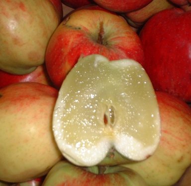
Figure 7. An apple with watercore: the glassy parts have liquid instead of air between the cells. Photo: Red58bill, CC BY 3.0, Wikimedia Commons.

So there is a way for an apple to reflect less without any new pigment: take away the air-filled boundaries between its cells. My guess said “fewer cells”; the physics says “fewer cell-to-air boundaries”. That is not what makes a cut apple brown, but it is a real way to make apple flesh darker, and I didn’t expect my wrong guess to land so close to it.
And real rot is someone else’s work, lol! The soft, watery, spreading brown patch on a stored apple is mostly fungi. The most important one is Penicillium expansum, which causes blue mould; it enters through fresh wounds like stem punctures, bruises, and even scratches from pickers’ fingernails, and makes lesions which are soft, watery, and light brown, later covered with bluish-green spores26. So decay is the apple being eaten by fungi, and browning is the apple’s own chemistry; in neither case are fewer cells the reason.
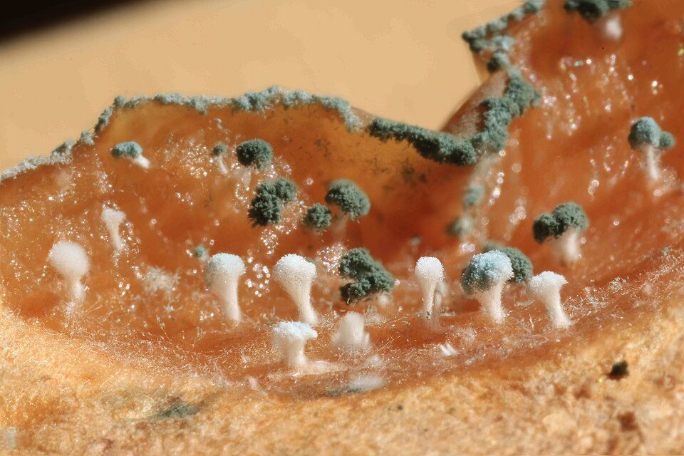
Figure 8. Blue mould on an apple; the little stalks carry the spores. Photo: ImagePerson, CC BY 4.0, Wikimedia Commons.

A red apple under yellow light
Now the fun one. Suppose the apple is pure red: it reflects only red light and absorbs everything else. Put it under a light source which emits only yellow. There is nothing in the incoming light which the apple can reflect, so nothing comes back, and the apple looks black.
Such a light source exists and was, for a long time, hanging over our streets: the low-pressure sodium lamp. Its light is almost entirely two lines very close to each other, at 588.995 nm and 589.592 nm27, the famous sodium D lines. Under this lamp there is “essentially no color rendition”28: every object looks like a brighter or darker shade of the same yellow-orange. (Astronomers love these lamps for exactly this reason: all the light pollution sits at one wavelength, which is easy to filter out28.)
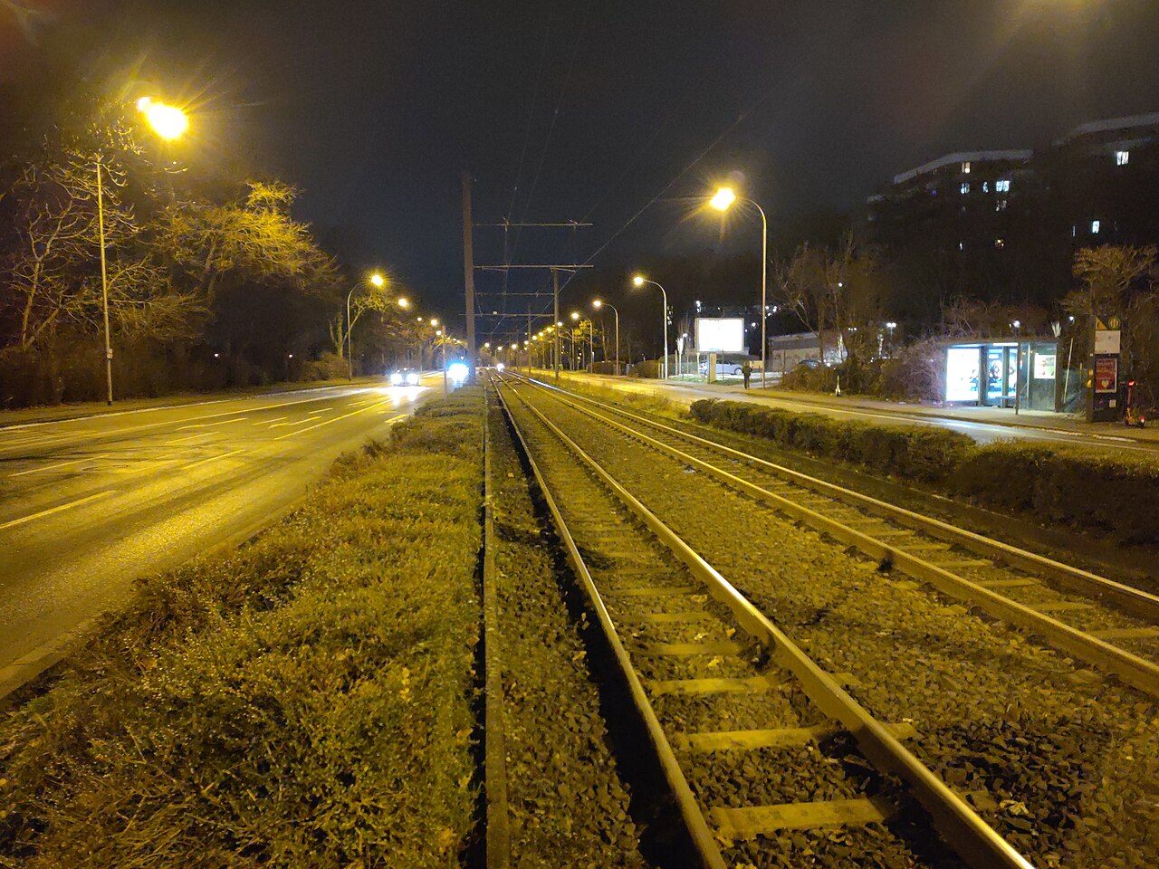
Figure 9. A street in Mainz, Germany, under low-pressure sodium lamps. The grass has no green left; only things with their own light, like the billboards, show colour. (The camera makes it look more yellow than it is to the eye.) Photo: ManuelB701, CC0, Wikimedia Commons.

A real apple is not pure red, so it is not perfectly black either. Look at the dashed line in Figure 4: at 589 nm the red apple sits right at the edge of its anthocyanin absorption, so it reflects only a little. Under a sodium lamp it would look like a dark, dull, brownish version of the lamp’s colour. A green apple reflects more at 589 nm, so it would likely look brighter than the red one. You would no longer be able to tell a red apple from a green apple by colour at all; only by how bright or dark it is.
Our eyes explain why. Look at the dashed line in Figure 2: at 589 nm the S-cones hardly respond at all, and the M- and L-cones respond in a fixed ratio. Whatever the object, the brain gets the same pair of numbers, just scaled up or down with brightness. One ratio means one colour, and all that is left is light and dark.
Don’t test this with your phone
Here is the trap. If you open a picture of plain yellow on your phone and hold an apple in front of it, the apple will still look reddish. Why? Because your screen has no yellow light. Each pixel has only red, green, and blue sub-pixels, and “yellow” on a screen is red plus green light at the same time. Our eyes can’t tell the difference: all that matters is how strongly the M- and L-cones fire, and a suitable mix of red and green makes them fire just like pure 589 nm light does. This is the whole reason almost any colour can be matched by mixing three primaries4. Two lights with completely different spectra look the same to us, but the apple is not fooled: it reflects the red part of your “yellow” screen and looks red.
So to do this experiment properly, you need pure monochromatic light:
- A sodium lamp, if you can still find one.
- A sodium flame. Sprinkle a pinch of table salt into a gas flame (a gas stove works) in a dark room. The bright yellow you see is the same sodium D-line emission27. It is not very bright, so keep the apple close, and be careful with the fire.
- A narrow-band filter around 589 nm in front of a white torch, if you happen to have one in a lab.
Put a red apple, a green apple, and something white next to each other and watch the colours drain out. You may send me photos at anantnug[at]gmail[dot]com if you try this :)
Try it yourself
If you don’t have a sodium lamp lying around (I don’t either), here is a small game. For every question, the page actually computes the colour: it multiplies the spectrum of the light with how much the object reflects at each wavelength, feeds that into the standard colour-matching functions of the human eye29, and converts the result into a colour your screen can show. Sunlight is the CIE standard daylight spectrum D6530, and the leaf is a real apple leaf, measured with a spectroradiometer31. Guess first, then read why.
About the game: the two apples use the sketched curves of Figure 4, not measured data. The phone’s “yellow” is two narrow bands at 530 and 620 nm, mixed so the cones see it like 589 nm light; real screens differ a little. White paper reflects 90% everywhere. And no screen can show the real sodium yellow, so you see the closest it can do.
So, how does anything take colour?
An object’s colour is the part of the light which it doesn’t absorb, as decoded by three types of cells in our eyes. The absorbing is done by molecules which behave like tiny tuned receivers, and the reflecting is helped along by every boundary where the refractive index jumps. An apple is green when chlorophyll is taking out the red and blue, red when anthocyanin is taking out the green, brown when broken cells let an enzyme build a new brown pigment, and glassy when its air pockets fill with liquid. Change the light source and the colour changes with it.
Until next time.
PS: I didn’t read as much chemistry in my high school as I read to write this article, but it was all worth it.
कुछ सवाल मुझे काफ़ी समय से परेशान कर रहे थे:
- किसी भी चीज़ को रंग मिलता कैसे है?
- जो सेब पूरा बड़ा नहीं हुआ, वो हरा दिखता है, और वक़्त के साथ उसका रंग बदल जाता है। कैसे?
- सेब सड़ता है तो काला पड़ जाता है। क्या ऐसा इसलिए है कि सेल टूट रहे हैं और रोशनी लौटाने के लिए कम सेल बचे हैं?
- अगर धूप में कोई सेब एकदम लाल है, तो वो ऐसी रोशनी में कैसा दिखेगा जिसमें लाल का नाम-ओ-निशान न हो, मान लो एक मोनोक्रोमैटिक (monochromatic) पीली रोशनी?
तीसरे सवाल पर मेरा अपना एक अंदाज़ा भी था, जिस पर मैं आगे लौटूँगा, क्योंकि वो एक दिलचस्प तरीक़े से ग़लत निकला।
मैं रोज़ इलेक्ट्रोमैग्नेटिक वेव्स (electromagnetic waves) के साथ काम करता हूँ, ज़्यादातर कुछ MHz से कुछ GHz के बीच। रोशनी भी वही चीज़ है, बस बहुत, बहुत ऊँची फ़्रीक्वेंसी पर। \(\lambda = 600\) nm वेवलेंथ (wavelength) वाली लाल रोशनी इतनी फ़्रीक्वेंसी पर दोलन करती है:
\[f = \frac{c}{\lambda} = \frac{3 \times 10^{8}\ \text{m/s}}{600 \times 10^{-9}\ \text{m}} = 5 \times 10^{14}\ \text{Hz},\]यानी 500 THz। तो, ये टॉपिक मेरे लिए बहुत अनजाना नहीं है, और एक अलग सेक्शन में मैं इसे वैसे ही देखूँगा जैसे अपने एंटीना और कॉइल्स को देखता हूँ।
किसी चीज़ को रंग मिलता कैसे है?
धूप में वो सारी वेवलेंथ होती हैं जो हमारी आँखें देख सकती हैं, आम तौर पर 380 से 700 nm तक1। जब ये रोशनी किसी चीज़ पर पड़ती है, तो उसके मॉलिक्यूल (molecule) कुछ वेवलेंथ एब्ज़ॉर्ब (absorb) कर लेते हैं और बाक़ी को वापस भेज देते हैं (या आर-पार जाने देते हैं)। जो कुछ वापस आकर हमारी आँख में जाता है, उसी को हम उस चीज़ का रंग कहते हैं।
पत्ता इसकी सबसे जानी-मानी मिसाल है। क्लोरोफ़िल (chlorophyll) ज़्यादातर नीले (400 से 500 nm) और लाल (650 से 680 nm) में एब्ज़ॉर्ब करता है, और हरे में, 530 nm के आस-पास, बस थोड़ा सा। तो हरी रोशनी पत्ते से बिखरकर बाहर लौट आती है, और पत्ता हरा दिखता है2। नीचे नापे गए स्पेक्ट्रम में ये सीधे दिखता है: दोनों क्लोरोफ़िल में दो कूबड़ हैं, एक नीले में और एक लाल में, और बीच में एक बड़ी घाटी, जो ठीक वही हरा है जो हम तक लौटता है।
चित्र 1. क्लोरोफ़िल a, क्लोरोफ़िल b और बीटा-कैरोटीन का नापा गया एब्ज़ॉर्प्शन, हर एक को उसकी अपनी पीक पर स्केल किया गया है। ये पत्ते के अंदर नहीं, एक सॉल्वेंट में नापे गए हैं। डेटा: PhotochemCAD3, OMLC के ज़रिए।
इस कहानी का दूसरा आधा हिस्सा हमारी आँखों में है। हमारे पास तीन तरह के कोन सेल (cone cells) हैं, जिनकी संवेदनशीलता क़रीब 440 nm (S-कोन), 545 nm (M-कोन) और 565 nm (L-कोन) पर पीक करती है4। दिमाग़ को पूरा स्पेक्ट्रम नहीं मिलता; उसे तीन नंबर मिलते हैं, यानी हर तरह का कोन कितनी ज़ोर से फ़ायर हुआ, और वो इन्हीं से रंग का एहसास गढ़ता है। ये बात याद रखना, आख़िरी सेक्शन में ये हमें काटने लौटेगी।
चित्र 2. हमारी आँख के तीनों तरह के कोन कितने संवेदनशील हैं, 1 की पीक पर स्केल किया गया। डेटा: Stockman और Sharpe5, CVRL डेटाबेस से। डैश वाली लाइन को अभी के लिए छोड़ दो।
थोड़ा और गहराई में: कोई मॉलिक्यूल सिर्फ़ कुछ ही रंग क्यों एब्ज़ॉर्ब करता है?
रोशनी तब एब्ज़ॉर्ब होती है जब उसकी एनर्जी ठीक उतनी हो जितनी मॉलिक्यूल के किसी इलेक्ट्रॉन को ऊपर के लेवल पर कूदने के लिए चाहिए। ज़्यादातर छोटे मॉलिक्यूल को इतनी ज़्यादा एनर्जी चाहिए कि वो सिर्फ़ अल्ट्रावायलेट एब्ज़ॉर्ब करते हैं, इसीलिए वो बेरंग दिखते हैं। पर जिन मॉलिक्यूल में बारी-बारी से सिंगल और डबल बॉन्ड की लंबी चेन होती है (इन्हें कॉन्जुगेटेड (conjugated) सिस्टम कहते हैं), उनमें इलेक्ट्रॉन पूरी चेन पर फैले रहते हैं। वो जितने ज़्यादा फैले (डीलोकलाइज़्ड) होते हैं, एनर्जी का गैप उतना छोटा होता है, और वो उतनी लंबी वेवलेंथ एब्ज़ॉर्ब करते हैं6। गाजर का पिगमेंट बीटा-कैरोटीन, जिसमें 11 कॉन्जुगेटेड डबल बॉन्ड हैं, अपना एब्ज़ॉर्प्शन नीले तक ले जाता है6; चित्र 1 में ये क़रीब 400 से 500 nm तक एब्ज़ॉर्ब करता है और उसके आगे कुछ नहीं, इसीलिए गाजर नारंगी दिखती है। क्लोरोफ़िल और सेब के लाल पिगमेंट, दोनों में ऐसी कॉन्जुगेटेड बनावट है, और ठीक इसी वजह से वो UV में नहीं, दिखने वाली रेंज में एब्ज़ॉर्ब करते हैं।
इलेक्ट्रोमैग्नेटिक नज़रिया
ऊपर जो कुछ है वो केमिस्ट्री है: इलेक्ट्रॉन, बॉन्ड, एनर्जी लेवल। पर रोशनी एक इलेक्ट्रोमैग्नेटिक वेव है, और मैं ये पूछे बिना नहीं रह सकता कि मेरा अपना फ़ील्ड इस सब के बारे में क्या कहता है। पता चला कि काफ़ी कुछ कहता है।
पहले, एनर्जी। \(\nu\) फ़्रीक्वेंसी वाला फ़ोटॉन \(E = h\nu = hc/\lambda\) एनर्जी लेकर चलता है, जहाँ \(h = 6.626\,070\,15 \times 10^{-34}\) J/Hz प्लैंक कॉन्स्टेंट (Planck constant) है7। 550 nm की हरी रोशनी के लिए,
\[E = \frac{hc}{\lambda} = \frac{(6.626 \times 10^{-34}\ \text{J s})(3 \times 10^{8}\ \text{m/s})}{550 \times 10^{-9}\ \text{m}} \approx 3.6 \times 10^{-19}\ \text{J} \approx 2.25\ \text{eV}.\]पूरी दिखने वाली रेंज, 380 से 700 nm, मोटे तौर पर 3.3 eV से 1.8 eV तक है। तो कोई मॉलिक्यूल रंगीन तभी होगा जब उसमें इलेक्ट्रॉन की कोई ऐसी छलाँग हो जिसकी क़ीमत 1.8 और 3.3 eV के बीच हो, और ऊपर वाले बॉक्स की कॉन्जुगेटेड चेन ठीक वही तरीक़ा हैं जिससे क़ुदरत गैप को इस खिड़की में ले आती है।
दूसरा, और ये वो हिस्सा है जो मुझे सबसे ज़्यादा पसंद है: पिगमेंट का मॉलिक्यूल एक नन्हे एंटीना की तरह बर्ताव करता है। किसी मटीरियल का सबसे सीधा क्लासिकल मॉडल, लॉरेंट्ज़ ऑसिलेटर मॉडल (Lorentz oscillator model), हर बँधे हुए इलेक्ट्रॉन को स्प्रिंग पर लगे एक मास की तरह देखता है8। स्प्रिंग है न्यूक्लियस का खिंचाव, जो इलेक्ट्रॉन को एक नैचुरल (रेज़ोनेंस) फ़्रीक्वेंसी \(\omega_0\) देता है। रोशनी की वेव का दोलन करता इलेक्ट्रिक फ़ील्ड इलेक्ट्रॉन को आगे-पीछे धकेलता है, और घर्षण जैसी डैम्पिंग \(\gamma\) इस हरकत से एनर्जी निकालती रहती है। तब इलेक्ट्रॉन के लिए न्यूटन का दूसरा नियम है8
\[\frac{d^2x}{dt^2} + \gamma \frac{dx}{dt} + \omega_0^2 x = \frac{qE_0}{m} \cos(\omega t),\]जो वही इक्वेशन है जो धकेली जा रही, डैम्प होती स्प्रिंग की, या धकेले जा रहे RLC सर्किट की होती है। इसे हल करके प्रति यूनिट वॉल्यूम ऐसे \(N\) इलेक्ट्रॉन के डाइपोल जोड़ो, तो मटीरियल की परमिटिविटी (permittivity) मिलती है8:
\[\varepsilon(\omega) = 1 + \frac{N q^2}{\varepsilon_0 m} \, \frac{1}{\omega_0^2 - \omega^2 - i\gamma\omega}.\]हर (denominator) में बैठा \(-i\gamma\omega\) \(\varepsilon\) को एक कॉम्प्लेक्स नंबर बना देता है, और इसलिए रिफ़्रैक्टिव इंडेक्स (refractive index) \(\tilde{n} = \sqrt{\varepsilon}\) भी कॉम्प्लेक्स हो जाता है। इसका वास्तविक हिस्सा तय करता है कि रोशनी कितनी मुड़ेगी (स्नेल का नियम), और काल्पनिक हिस्सा तय करता है कि कितनी रोशनी एब्ज़ॉर्ब होगी8। चित्र 3 दिखाता है कि रेज़ोनेंस के आस-पास ये दोनों हिस्से कैसे दिखते हैं।
चित्र 3. \(Q = 8\) वाला एक लॉरेंट्ज़ ऑसिलेटर, ऊपर वाली इक्वेशन से प्लॉट किया गया, तो ये मॉडल है, कोई माप नहीं। एब्ज़ॉर्प्शन (लाल) रेज़ोनेंस पर पीक करता है; वास्तविक हिस्सा (नीला, डैश वाला) उसके आर-पार साइन बदल लेता है।
अब इसे एक इंजीनियर की नज़र से देखो। वायरलेस पावर ट्रांसफ़र में रिसीवर कॉइल को ऐसे ट्यून किया जाता है कि वो ट्रांसमीटर की फ़्रीक्वेंसी पर रेज़ोनेट करे; रेज़ोनेंस पर वो फ़ील्ड से सबसे ज़्यादा पावर खींचती है, और रेज़ोनेंस से थोड़ा हटते ही बहुत कम। ये खिड़की कितनी चौड़ी है, ये लॉस पर निर्भर करता है, यानी क्वालिटी फ़ैक्टर (quality factor) \(Q = \omega_0 / \gamma\) पर। पिगमेंट 500 THz पर यही चीज़ है: क्लोरोफ़िल के रेज़ोनेंस नीले और लाल में हैं, और यही वो फ़्रीक्वेंसी हैं जो वो धूप से निकाल लेता है (चित्र 1)। चित्र 1 के चौड़े कूबड़, इस भाषा में, कम-\(Q\) वाले रेज़ोनेंस हैं। तो जब मैं कहता हूँ कि सेब “हरा एब्ज़ॉर्ब करता है”, तो असल में मेरा मतलब है कि सेब का छिलका हरे पर ट्यून किए गए नन्हे रिसीवरों से भरा है।
यही मॉडल ये भी बताता है कि किसी बाउंड्री पर क्या होता है, जो सड़ने वाले सेक्शन में काम आएगा। दो मटीरियल की बाउंड्री पर सीधी पड़ती रोशनी के लिए, लौटने वाली पावर का हिस्सा फ़्रेनेल इक्वेशन (Fresnel equation) देती है8
\[R = \left| \frac{\tilde{n}_1 - \tilde{n}_2}{\tilde{n}_1 + \tilde{n}_2} \right|^2 .\]रिफ़्रैक्टिव इंडेक्स में फ़र्क़ नहीं, तो रिफ़्लेक्शन नहीं। इसे याद रखना।
हरा सेब लाल क्यों हो जाता है?
कच्चा सेब उसी वजह से हरा होता है जिस वजह से पत्ता: उसका छिलका क्लोरोफ़िल से भरा होता है। जैसे-जैसे सेब पकता है, छिलके में एक साथ दो चीज़ें होती हैं।
पहली, हरा चला जाता है। पकते वक़्त क्लोरोफ़िल टूटकर फ़ाइलोबिलिन (phyllobilins) नाम के बेरंग कंपाउंड में बदल जाता है; ‘Gala’ सेबों के छिलके में उनकी शेल्फ़ लाइफ़ के दौरान इसे ट्रैक किया गया है9। किसानों के लिए लिखी एक्सटेंशन गाइड यही बात बाहर से देखकर बताती हैं: फल पकता है तो “हरा बैकग्राउंड रंग फीका पड़ जाता है”10।
दूसरी, लाल बनता है। उसी वक़्त छिलके के सेल एंथोसायनिन (anthocyanins) बनाने लगते हैं, पिगमेंट का वही परिवार जो लाल पत्तागोभी और ब्लूबेरी को रंग देता है। सेब के छिलके में एक एंथोसायनिन, सायनिडिन 3-गैलेक्टोसाइड, कुल का 85% से ज़्यादा होता है11। सेब के छिलके से लौटी रोशनी के माप दिखाते हैं कि एंथोसायनिन 550 nm के पास एब्ज़ॉर्ब करते हैं12, यानी हरे में। पिछले सेक्शन की एंटीना वाली भाषा में, सेब अपने रिसीवर बदल लेता है: नीले और लाल पर ट्यून वाले उतार दिए जाते हैं, और हरे पर ट्यून वाले नए लगा दिए जाते हैं। धूप से हरा निकाल दो, तो जो लौटता है वो ज़्यादातर लाल है।
नीचे वाला चित्र एक स्केच है कि स्पेक्ट्रम के रूप में ये कैसा दिखता है।
चित्र 4. सांकेतिक, नापा हुआ डेटा नहीं। मुझे सेब के छिलके का खुले लाइसेंस वाला नापा हुआ स्पेक्ट्रम नहीं मिला, तो मैंने Merzlyak et al.12 के एब्ज़ॉर्प्शन बैंड से शेप बनाए। डैश वाली लाइन को अभी के लिए छोड़ दो।
सेब लाल बनाना कब शुरू करेगा, ये कौन तय करता है? पता चला कि एक मास्टर स्विच है: MdMYB10 नाम का एक जीन। ये एक प्रोटीन बनाता है जो एंथोसायनिन बनाने के लिए ज़रूरी सारे जीन चालू कर देता है। इसकी एक्टिविटी ठीक तब बढ़ती है जब रंग बनता है, और जिन सेब के पौधों को इसे ज़्यादा बनाने के लिए इंजीनियर किया गया, वो ख़ूब रंगीन हो गए13। इस स्विच के बारे में मैंने जो दो सबसे दिलचस्प बातें सीखीं, वो मौसम से जुड़ी हैं:
- इसे धूप चाहिए। अँधेरे में कोई एंथोसायनिन नहीं बनता11, और ज़्यादा धूप से छिलके में ज़्यादा पिगमेंट बनता है, एक हद तक14। शायद इसीलिए सेब का धूप की तरफ़ वाला हिस्सा पेड़ की तरफ़ वाले हिस्से से अक्सर ज़्यादा लाल होता है।
- इसे ठंडी रातें पसंद हैं। पेड़ पर लगे सेबों को गरम करने से पिगमेंट और MYB10 की एक्टिविटी, दोनों बहुत घट गए, जबकि कम तापमान की एक ही रात MYB10 को बड़ा बढ़ावा देने के लिए काफ़ी थी15। इसीलिए गरम इलाक़ों में उगे सेबों में रंग ठीक से नहीं आता। हिमाचल में ये दिखता है। निचली पहाड़ियों के पुराने Delicious सेबों में रंग कम आता है, और सरकार के अपने एक्सटेंशन सेंटर अब कम ऊँचाई पर कम रंग को ऐसी दिक़्क़त बताते हैं जिसे क्लाइमेट चेंज और बढ़ा रहा है; रोहड़ू के आस-पास की बीच वाली पहाड़ियों में बाग़वान इन्हें ‘Red Chief’ और ‘Scarlet Spur’ जैसी ज़्यादा रंग वाली किस्मों से बदल रहे हैं16। बाग़ ख़ुद भी ऊपर चढ़ रहे हैं: 1980 के दशक में सेब 1200 से 1500 m के बीच उगाए जाते थे, 2000 के दशक में 1500 से 2500 m के बीच, और 2014 तक 3500 m से ऊपर, ऊपरी किन्नौर और लाहौल-स्पीति में, क्योंकि नीचे की घाटियाँ गरम होती गईं17।
और अगर सेब पीला है, जैसे Golden Delicious, तो क्लोरोफ़िल तब भी जाता है पर एंथोसायनिन बहुत कम बनता है, तो पीले कैरोटिनॉयड, जो इतने समय से हरे के नीचे छिपे बैठे थे, दिखने लगते हैं (सेब के छिलके का रंग सिर्फ़ एंथोसायनिन पर नहीं, क्लोरोफ़िल और कैरोटिनॉयड पर भी निर्भर करता है11)।
एक और बात, जो अगले सेक्शन के लिए ज़रूरी है: तोड़ लेने के बाद भी सेब पकना बंद नहीं करता। सेब एक क्लाइमैक्टेरिक (climacteric) फल है। ऐसे फल तुड़ाई के बाद भी पक सकते हैं, और पकते वक़्त वो तेज़ साँस लेते हैं और एथिलीन (ethylene) का एक झोंका छोड़ते हैं, एक गैस जो पकाने वाले हॉर्मोन का काम करती है और अपने आप को और बनवाती है10। कुछ किस्में इतनी ज़्यादा एथिलीन बनाती हैं कि स्टोरेज में जल्दी नरम पड़ जाती हैं और बूढ़ी हो जाती हैं18। तो तुम्हारी फल की टोकरी में रखा सेब अच्छा-ख़ासा ज़िंदा है।
सड़ने के बारे में मेरा अंदाज़ा, और वो ग़लत क्यों है
मेरा अंदाज़ा ये था:
अगर लगातार केमिकल मेल-जोल चल रहे हैं और पेड़ से तोड़ते ही सेल टूटने लगते हैं, तो रोशनी लौटाने के लिए कम सेल बचेंगे, इसलिए फल काला पड़ता है, जिसे हम सड़ना कहते हैं, यानी ज़्यादा एब्ज़ॉर्प्शन और कम रिफ़्लेक्शन।
सुनने में ठीक लगता है। आख़िरी हिस्सा तो सही भी है: भूरा सेब सच में ज़्यादा एब्ज़ॉर्ब करता है और कम लौटाता है। पर वजह ये नहीं कि सेल ग़ायब हो जाते हैं। क्यों, ये रहा।
पहली बात, टाइम स्केल मेल नहीं खाता। सेब काटकर मेज़ पर छोड़ दो: सतह मिनटों में भूरी हो जाती है। कुछ मिनटों में कोई ख़ास तादाद में सेल ग़ायब नहीं होते, तो ज़रूर कुछ जुड़ रहा है।
और जो जुड़ता है वो एक नया पिगमेंट है। सेब के सेल के अंदर पॉलीफ़ीनॉल ऑक्सीडेज़ (polyphenol oxidase, PPO) एंज़ाइम प्लास्टिड (plastids) नाम के छोटे डिब्बों में रहता है, जबकि उसका कच्चा माल, फ़ीनॉलिक कंपाउंड, वैक्यूओल (vacuole) में रखा रहता है। स्वस्थ सेल में दोनों कभी मिलते नहीं, तो भूरापन नहीं आता। जब ये डिब्बे टूटते हैं, काटने से, दबने से, कीड़ों से, या बस उम्र से, तो एंज़ाइम ऑक्सीजन की मौजूदगी में फ़ीनॉलिक्स से मिलता है और उन्हें क्विनोन (quinones) में बदल देता है, जो फिर आपस में जुड़कर भूरे पॉलिमर बना लेते हैं19। ताज़ा कटे सेब, प्यूरी और जूस के भूरे पड़ने के पीछे भी यही तरीक़ा है, जिसे रोकने में फ़ूड इंडस्ट्री काफ़ी मेहनत करती है20। इलेक्ट्रोमैग्नेटिज़्म की भाषा में, टूटा सेल रिसीवरों का एक बिल्कुल नया सेट खड़ा कर लेता है।
चित्र 5. एंज़ाइम से होने वाला भूरापन, Murata19 के मुताबिक़।
सबसे अच्छा सबूत है एक सेब जो भूरा नहीं होता। Arctic सेब जेनेटिकली इंजीनियर किया गया एक सेब है जिसमें चार PPO जीन दबा दिए गए हैं, और नतीजा है एक ऐसा सेब जो भूरा नहीं पड़ता। अगर भूरापन सेलों के ग़ायब होने और कम लौटाने की वजह से होता, तो एक एंज़ाइम को चुप कराने से वो रुकता नहीं।
जहाँ मेरा अंदाज़ा सोच से ज़्यादा क़रीब था
एक जगह है जहाँ “कम रिफ़्लेक्शन” बिना किसी नए पिगमेंट के होता है, और वो पूरी तरह इलेक्ट्रोमैग्नेटिक्स है।
सोचो, सेब का गूदा सफ़ेद होता ही क्यों है। उसमें लगभग कोई पिगमेंट नहीं; सेब का कॉर्टेक्स आम तौर पर बेरंग होता है13। पानी का गिलास भी बेरंग होता है, पर वो पारदर्शी है, सफ़ेद नहीं। फ़र्क़ बनावट का है। सेब का गूदा कोई ठोस टुकड़ा नहीं है: किस्म के हिसाब से सेब के वॉल्यूम का 30% तक हवा हो सकती है, जो सेलों के बीच की जगहों में बैठी रहती है21। ‘Jonagold’ सेबों में गूदे का हवा वाला हिस्सा सीज़न की शुरुआत के 10.6% से बढ़कर तुड़ाई तक 26.4% हो गया21। तो गूदे में जाती रोशनी की एक किरण सेल वॉल, हवा, सेल वॉल, हवा, बार-बार पार करती है, और हर बार पिछले सेक्शन वाली फ़्रेनेल इक्वेशन थोड़ा सा हिस्सा वापस भेज देती है।
कितना? पौधों की सेल वॉल का रिफ़्रैक्टिव इंडेक्स क़रीब 1.425 है22, पानी का क़रीब 1.3323, और हवा का 1.0। इन्हें फ़्रेनेल इक्वेशन में डालो:
\[R_{\text{wall|air}} = \left(\frac{1.425 - 1}{1.425 + 1}\right)^2 \approx 3.1\%,\] \[R_{\text{wall|water}} = \left(\frac{1.425 - 1.33}{1.425 + 1.33}\right)^2 \approx 0.12\%.\]चित्र 6. एक बाउंड्री पर लौटी रोशनी, फ़्रेनेल इक्वेशन से, सेल वॉल का रिफ़्रैक्टिव इंडेक्स 1.42522 और पानी का 1.3323 लेकर।
एक बाउंड्री सिर्फ़ 3% लौटाती है, पर गूदे के अंदर रोशनी ऐसी बहुत सारी बाउंड्री से टकराती है, और थोड़ा-थोड़ा करके उसका ज़्यादातर हिस्सा बाहर वापस आ जाता है, तो गूदा सफ़ेद दिखता है। अब हवा की जगह पानी भर दो: हर बाउंड्री क़रीब 25 गुना कम लौटाती है, रोशनी गहराई तक जाती है और एब्ज़ॉर्ब हो जाती है या आर-पार निकल जाती है, और टिश्यू ज़्यादा गहरा और काँच जैसा दिखता है। ये सिर्फ़ हिसाब नहीं है। जब पत्ते की हवा वाली जगहों में तरल भर दिया जाता है, तो पत्ता पूरी 500 से 2500 nm रेंज में कम रोशनी लौटाता है, ठीक इसलिए कि सेल वॉल और हवा वाली बाउंड्री हट जाती हैं2224। और सेब ये अपने साथ ख़ुद करते हैं। वॉटरकोर (watercore) नाम की एक गड़बड़ी में गूदे की हवा वाली जगहें सॉर्बिटॉल से भरे तरल से भर जाती हैं, और वो गूदा पारभासी और काँच जैसा दिखता है25।
चित्र 7. वॉटरकोर वाला एक सेब: काँच जैसे हिस्सों में सेलों के बीच हवा की जगह तरल है। फ़ोटो: Red58bill, CC BY 3.0, Wikimedia Commons।
तो सेब के लिए बिना किसी नए पिगमेंट के कम लौटाने का एक तरीक़ा है: उसके सेलों के बीच की हवा भरी बाउंड्री हटा दो। मेरे अंदाज़े ने कहा “कम सेल”; फ़िज़िक्स कहती है “सेल और हवा के बीच की कम बाउंड्री”। कटा सेब इस वजह से भूरा नहीं होता, पर ये सेब के गूदे को गहरा करने का एक असली तरीक़ा है, और मैंने नहीं सोचा था कि मेरा ग़लत अंदाज़ा इसके इतने क़रीब जा गिरेगा।
और असली सड़न किसी और का काम है, lol! स्टोर किए सेब पर फैलता नरम, पानी जैसा, भूरा धब्बा ज़्यादातर फ़ंगस है। सबसे अहम है Penicillium expansum, जो ब्लू मोल्ड (blue mould) करता है; ये ताज़ा घावों से अंदर जाता है, जैसे डंठल के छेद, चोट, और तोड़ने वालों के नाख़ूनों की खरोंच तक, और ऐसे धब्बे बनाता है जो नरम, पानी जैसे और हल्के भूरे होते हैं, और बाद में नीले-हरे स्पोर से ढक जाते हैं26। तो सड़ना है सेब को फ़ंगस का खा जाना, और भूरापन है सेब की अपनी केमिस्ट्री; दोनों में से किसी में भी वजह कम सेल नहीं हैं।
चित्र 8. सेब पर ब्लू मोल्ड; छोटी डंडियों पर स्पोर लगे हैं। फ़ोटो: ImagePerson, CC BY 4.0, Wikimedia Commons।
पीली रोशनी में लाल सेब
अब मज़े वाला सवाल। मान लो सेब एकदम लाल है: वो सिर्फ़ लाल रोशनी लौटाता है और बाक़ी सब एब्ज़ॉर्ब कर लेता है। उसे ऐसी रोशनी में रखो जो सिर्फ़ पीला देती है। आने वाली रोशनी में ऐसा कुछ है ही नहीं जिसे सेब लौटा सके, तो कुछ वापस नहीं आता, और सेब काला दिखता है।
ऐसी रोशनी मौजूद है और बहुत समय तक हमारी सड़कों पर लटकी रही: लो-प्रेशर सोडियम लैंप। इसकी रोशनी लगभग पूरी की पूरी दो लाइनें हैं जो एक-दूसरे के बहुत पास हैं, 588.995 nm और 589.592 nm पर27, मशहूर सोडियम D लाइनें। इस लैंप के नीचे “रंग लगभग बिल्कुल नहीं दिखते”28: हर चीज़ उसी पीले-नारंगी का हल्का या गहरा शेड लगती है। (एस्ट्रोनॉमर्स को ये लैंप ठीक इसी वजह से पसंद हैं: सारा लाइट पॉल्यूशन एक ही वेवलेंथ पर होता है, जिसे फ़िल्टर करना आसान है28।)
चित्र 9. जर्मनी के माइंज़ शहर की एक सड़क, लो-प्रेशर सोडियम लैंप के नीचे। घास में कोई हरा नहीं बचा; सिर्फ़ अपनी रोशनी वाली चीज़ें, जैसे बिलबोर्ड, रंग दिखाती हैं। (कैमरा इसे आँख से दिखने के मुक़ाबले ज़्यादा पीला दिखाता है।) फ़ोटो: ManuelB701, CC0, Wikimedia Commons।
असली सेब एकदम लाल नहीं होता, तो वो पूरा काला भी नहीं दिखता। चित्र 4 की डैश वाली लाइन देखो: 589 nm पर लाल सेब अपने एंथोसायनिन एब्ज़ॉर्प्शन के ठीक किनारे पर है, तो वो बस थोड़ा सा लौटाता है। सोडियम लैंप के नीचे वो लैंप के रंग का एक गहरा, फीका, भूरा सा रूप दिखेगा। हरा सेब 589 nm पर ज़्यादा लौटाता है, तो शायद वो लाल वाले से ज़्यादा चमकीला दिखेगा। तुम लाल सेब को हरे सेब से रंग के आधार पर बिल्कुल अलग नहीं कर पाओगे; सिर्फ़ इससे कि वो कितना चमकीला या गहरा है।
हमारी आँखें बताती हैं क्यों। चित्र 2 की डैश वाली लाइन देखो: 589 nm पर S-कोन लगभग बिल्कुल रिस्पॉन्ड नहीं करते, और M- और L-कोन एक तय अनुपात में रिस्पॉन्ड करते हैं। चीज़ कोई भी हो, दिमाग़ को नंबरों की वही जोड़ी मिलती है, बस चमक के हिसाब से छोटी या बड़ी। एक अनुपात मतलब एक रंग, और बचता है सिर्फ़ हल्का और गहरा।
इसे अपने फ़ोन से मत आज़माना
यहीं फँसने वाली बात है। अगर तुम अपने फ़ोन पर सादे पीले रंग की तस्वीर खोलो और उसके सामने सेब पकड़ो, तो सेब फिर भी लाल सा दिखेगा। क्यों? क्योंकि तुम्हारी स्क्रीन में पीली रोशनी है ही नहीं। हर पिक्सल में सिर्फ़ लाल, हरे और नीले सब-पिक्सल होते हैं, और स्क्रीन का “पीला” एक साथ लाल और हरी रोशनी है। हमारी आँखें फ़र्क़ नहीं बता सकतीं: मायने बस इतना रखता है कि M- और L-कोन कितनी ज़ोर से फ़ायर होते हैं, और लाल और हरे का सही मेल उन्हें ठीक वैसे ही फ़ायर करवाता है जैसे शुद्ध 589 nm की रोशनी। यही पूरी वजह है कि लगभग कोई भी रंग तीन प्राइमरी रंग मिलाकर बनाया जा सकता है4। बिल्कुल अलग स्पेक्ट्रम वाली दो रोशनियाँ हमें एक जैसी दिखती हैं, पर सेब धोखा नहीं खाता: वो तुम्हारी “पीली” स्क्रीन का लाल हिस्सा लौटाता है और लाल दिखता है।
तो ये एक्सपेरिमेंट ठीक से करने के लिए शुद्ध मोनोक्रोमैटिक रोशनी चाहिए:
- सोडियम लैंप, अगर अब भी कहीं मिल जाए।
- सोडियम की लौ। अँधेरे कमरे में गैस की लौ में (गैस स्टोव चलेगा) एक चुटकी नमक छिड़को। जो चमकीला पीला दिखता है, वो वही सोडियम D-लाइन वाली रोशनी है27। ये बहुत तेज़ नहीं होती, तो सेब पास रखना, और आग से सावधान रहना।
- सफ़ेद टॉर्च के आगे 589 nm के आस-पास का एक नैरो-बैंड फ़िल्टर, अगर लैब में कहीं पड़ा हो।
एक लाल सेब, एक हरा सेब, और कोई सफ़ेद चीज़ साथ-साथ रखो और रंगों को बहते हुए देखो। अगर आज़माओ तो फ़ोटो मुझे anantnug[at]gmail[dot]com पर भेज सकते हो :)
ख़ुद आज़माओ
अगर तुम्हारे पास भी सोडियम लैंप नहीं पड़ा है (मेरे पास भी नहीं है), तो ये रहा एक छोटा सा गेम। हर सवाल के लिए पेज सच में रंग का हिसाब लगाता है: वो रोशनी के स्पेक्ट्रम को इससे गुणा करता है कि चीज़ हर वेवलेंथ पर कितना लौटाती है, उसे इंसानी आँख के स्टैंडर्ड कलर-मैचिंग फ़ंक्शन में डालता है29, और नतीजे को ऐसे रंग में बदलता है जो तुम्हारी स्क्रीन दिखा सके। धूप है CIE का स्टैंडर्ड डेलाइट स्पेक्ट्रम D6530, और पत्ता सेब का एक असली पत्ता है, जिसे स्पेक्ट्रोरेडियोमीटर से नापा गया है31। पहले अंदाज़ा लगाओ, फिर पढ़ो क्यों।
गेम के बारे में: दोनों सेब चित्र 4 के स्केच वाले कर्व इस्तेमाल करते हैं, नापा हुआ डेटा नहीं। फ़ोन का “पीला” 530 और 620 nm पर दो पतले बैंड हैं, ऐसे मिलाए गए कि कोन उसे 589 nm की रोशनी जैसा देखें; असली स्क्रीन थोड़ी अलग होती हैं। सफ़ेद काग़ज़ हर जगह 90% लौटाता है। और कोई भी स्क्रीन असली सोडियम वाला पीला नहीं दिखा सकती, तो तुम्हें उसके सबसे क़रीब वाला रंग दिखता है।
तो, किसी चीज़ को रंग मिलता कैसे है?
किसी चीज़ का रंग रोशनी का वो हिस्सा है जो वो एब्ज़ॉर्ब नहीं करती, जिसे हमारी आँखों के तीन तरह के सेल पढ़ते हैं। एब्ज़ॉर्ब करने का काम मॉलिक्यूल करते हैं जो नन्हे ट्यून किए गए रिसीवरों की तरह बर्ताव करते हैं, और लौटाने में हर वो बाउंड्री मदद करती है जहाँ रिफ़्रैक्टिव इंडेक्स उछलता है। सेब तब हरा है जब क्लोरोफ़िल लाल और नीला निकाल रहा है, तब लाल जब एंथोसायनिन हरा निकाल रहा है, तब भूरा जब टूटे सेल एक एंज़ाइम को नया भूरा पिगमेंट बनाने देते हैं, और तब काँच जैसा जब उसकी हवा वाली जेबें तरल से भर जाती हैं। रोशनी बदलो, तो रंग भी उसके साथ बदल जाता है।
फिर मिलेंगे।
PS: स्कूल में मैंने इतनी केमिस्ट्री नहीं पढ़ी थी जितनी ये आर्टिकल लिखने के लिए पढ़ी, पर ये सब वसूल था।
References
-
van Grondelle & Boeker, “Limits on Natural Photosynthesis”, J. Phys. Chem. B, 2017. link ↩
-
Dixon, Taniguchi & Lindsey, “PhotochemCAD 2”, Photochemistry and Photobiology, 2005. doi. Data files by S. Prahl, OMLC: link ↩
-
Stockman & Sharpe, “The spectral sensitivities of the middle- and long-wavelength-sensitive cones…”, Vision Research, 2000. doi. Data from CVRL: link ↩
-
J. Clark, “What Causes Molecules to Absorb UV and Visible Light”, Chemistry LibreTexts. link ↩ ↩2
-
J. Colton, “Lorentz Oscillator Model of the Dielectric Function”, BYU Physics 581 notes, 2020. pdf ↩ ↩2 ↩3 ↩4 ↩5
-
Gorfer et al., “Chlorophyll breakdown during fruit ripening… ‘Gala’…”, Food Research International, 2022. doi ↩
-
Capino & Farcuh, “Ethylene and the Regulation of Fruit Ripening”, University of Maryland Extension. link ↩ ↩2
-
Honda & Moriya, “Anthocyanin Biosynthesis in Apple Fruit”, Horticulture Journal, 2018. doi ↩ ↩2 ↩3
-
Merzlyak, Solovchenko & Gitelson, “Reflectance spectral features and non-destructive estimation of chlorophyll, carotenoid and anthocyanin content in apple fruit”, Postharvest Biology and Technology, 2003. doi ↩ ↩2
-
Espley et al., “Red colouration in apple fruit is due to the activity of the MYB transcription factor, MdMYB10”, The Plant Journal, 2007. doi ↩ ↩2
-
Chen et al., “Differential Regulation of Anthocyanin Synthesis in Apple Peel under Different Sunlight Intensities”, IJMS, 2019. doi ↩
-
Lin-Wang et al., “High temperature reduces apple fruit colour via modulation of the anthocyanin regulatory complex”, Plant, Cell & Environment, 2011. doi ↩
-
ICAR, “Coloured Strains of Apple for Enhancing Income in Mid-Hills under Climate Change Scenario” (KVK Rohru). link ↩
-
Sahu et al., “Why apple orchards are shifting to the higher altitudes of the Himalayas?”, PLoS ONE, 2020. doi ↩
-
University of Maine Extension, “The Role of Ethylene in Fruit Ripening”. link ↩
-
M. Murata, “Food chemistry and biochemistry of enzymatic browning”, Food Science and Technology Research, 2022. doi ↩ ↩2
-
Arnold & Gramza-Michałowska, “Enzymatic browning in apple products and its inhibition treatments: A comprehensive review”, CRFSFS, 2022. doi ↩
-
Herremans et al., “Spatial development of transport structures in apple fruit”, Frontiers in Plant Science, 2015. doi ↩ ↩2
-
Gausman, Allen & Escobar, “Refractive Index of Plant Cell Walls”, Applied Optics, 1974. doi ↩ ↩2 ↩3
-
Hale & Querry, “Optical Constants of Water in the 200-nm to 200-μm Wavelength Region”, Applied Optics, 1973. doi ↩ ↩2
-
J. T. Woolley, “Reflectance and Transmittance of Light by Leaves”, Plant Physiology, 1971. doi ↩
-
M. Farcuh, “Water core in apples”, University of Maryland Extension. link ↩
-
Janisiewicz & Biggs, “Blue Mold on Apple”, Extension Foundation. link ↩
-
NIST, “Strong Lines of Sodium”, Handbook of Basic Atomic Spectroscopic Data. link ↩ ↩2
-
Flagstaff Dark Skies Coalition, “Low Pressure Sodium Lighting”. link ↩ ↩2
-
CIE 1931 colour-matching functions, 2 degree observer. doi. Values taken from CVRL: link ↩
-
Braun et al., “A Multi-Temporal Field Spectroscopy Dataset of Apple Leaf Reflectance for Tree Vitality Monitoring”, Zenodo, 2026, CC BY 4.0. doi. The game uses the mean of the 30 spectra measured on 16 July 2025. ↩
संदर्भ
-
van Grondelle & Boeker, “Limits on Natural Photosynthesis”, J. Phys. Chem. B, 2017. link ↩
-
Dixon, Taniguchi & Lindsey, “PhotochemCAD 2”, Photochemistry and Photobiology, 2005. doi. Data files by S. Prahl, OMLC: link ↩
-
Stockman & Sharpe, “The spectral sensitivities of the middle- and long-wavelength-sensitive cones…”, Vision Research, 2000. doi. Data from CVRL: link ↩
-
J. Clark, “What Causes Molecules to Absorb UV and Visible Light”, Chemistry LibreTexts. link ↩ ↩2
-
J. Colton, “Lorentz Oscillator Model of the Dielectric Function”, BYU Physics 581 notes, 2020. pdf ↩ ↩2 ↩3 ↩4 ↩5
-
Gorfer et al., “Chlorophyll breakdown during fruit ripening… ‘Gala’…”, Food Research International, 2022. doi ↩
-
Capino & Farcuh, “Ethylene and the Regulation of Fruit Ripening”, University of Maryland Extension. link ↩ ↩2
-
Honda & Moriya, “Anthocyanin Biosynthesis in Apple Fruit”, Horticulture Journal, 2018. doi ↩ ↩2 ↩3
-
Merzlyak, Solovchenko & Gitelson, “Reflectance spectral features and non-destructive estimation of chlorophyll, carotenoid and anthocyanin content in apple fruit”, Postharvest Biology and Technology, 2003. doi ↩ ↩2
-
Espley et al., “Red colouration in apple fruit is due to the activity of the MYB transcription factor, MdMYB10”, The Plant Journal, 2007. doi ↩ ↩2
-
Chen et al., “Differential Regulation of Anthocyanin Synthesis in Apple Peel under Different Sunlight Intensities”, IJMS, 2019. doi ↩
-
Lin-Wang et al., “High temperature reduces apple fruit colour via modulation of the anthocyanin regulatory complex”, Plant, Cell & Environment, 2011. doi ↩
-
ICAR, “Coloured Strains of Apple for Enhancing Income in Mid-Hills under Climate Change Scenario” (KVK Rohru). link ↩
-
Sahu et al., “Why apple orchards are shifting to the higher altitudes of the Himalayas?”, PLoS ONE, 2020. doi ↩
-
University of Maine Extension, “The Role of Ethylene in Fruit Ripening”. link ↩
-
M. Murata, “Food chemistry and biochemistry of enzymatic browning”, Food Science and Technology Research, 2022. doi ↩ ↩2
-
Arnold & Gramza-Michałowska, “Enzymatic browning in apple products and its inhibition treatments: A comprehensive review”, CRFSFS, 2022. doi ↩
-
Herremans et al., “Spatial development of transport structures in apple fruit”, Frontiers in Plant Science, 2015. doi ↩ ↩2
-
Gausman, Allen & Escobar, “Refractive Index of Plant Cell Walls”, Applied Optics, 1974. doi ↩ ↩2 ↩3
-
Hale & Querry, “Optical Constants of Water in the 200-nm to 200-μm Wavelength Region”, Applied Optics, 1973. doi ↩ ↩2
-
J. T. Woolley, “Reflectance and Transmittance of Light by Leaves”, Plant Physiology, 1971. doi ↩
-
M. Farcuh, “Water core in apples”, University of Maryland Extension. link ↩
-
Janisiewicz & Biggs, “Blue Mold on Apple”, Extension Foundation. link ↩
-
NIST, “Strong Lines of Sodium”, Handbook of Basic Atomic Spectroscopic Data. link ↩ ↩2
-
Flagstaff Dark Skies Coalition, “Low Pressure Sodium Lighting”. link ↩ ↩2
-
CIE 1931 colour-matching functions, 2 degree observer. doi. Values taken from CVRL: link ↩
-
Braun et al., “A Multi-Temporal Field Spectroscopy Dataset of Apple Leaf Reflectance for Tree Vitality Monitoring”, Zenodo, 2026, CC BY 4.0. doi. The game uses the mean of the 30 spectra measured on 16 July 2025. ↩